File list
This special page shows all uploaded files.
| Date | Name | Thumbnail | Size | Description | Versions |
|---|---|---|---|---|---|
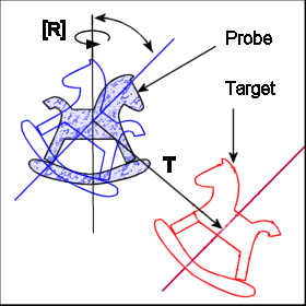
| 16:20, 8 April 2008 | RTfunctioninMR.gif (file) |  |
13 KB | 2 | |
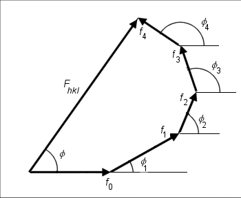
| 15:22, 1 April 2008 | Sfac4.gif (file) |  |
6 KB | Vector phase diagram showing the net diffracted wave scattered from a primitive unit cell containing atoms at 5 distinct positions. | 1 |
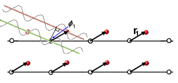
| 15:13, 1 April 2008 | Sfac2.gif (file) |  |
7 KB | 2 | |
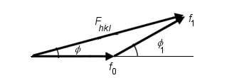
| 15:13, 1 April 2008 | Sfac3.gif (file) |  |
3 KB | 2 | |
| 14:46, 1 April 2008 | Sfac1.gif (file) |  |
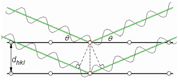
11 KB | X-ray diffraction from an array of atoms situated at lattice points in a primitive crystal. | 1 |
| 10:43, 1 April 2008 | Scatfacs.gif (file) |  |
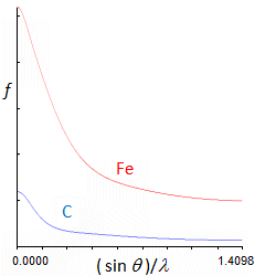
6 KB | Scattering factor of stationary C and Fe atoms plotted as a function of Bragg angle for incident X-ray wavelength of 0.70930 angstroms. Ticks on the horizontal axis correspond to Bragg angle increments of 10 degrees; ticks on the vertical axis are increme | 1 |
| 10:50, 2 March 2006 | Coverill.gif (file) |  |
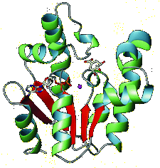
19 KB | Crystal structure of shikimate kinase from Mycobacterium tuberculosis (MtSK) complexed with MgADP and shikimic acid (shikimate) has been determined at 2.3 Å resolution, clearly revealing the amino-acid residues involved in shikimate binding. This is the | 1 |
| 14:54, 25 January 2006 | ReciprocalLattice-3.gif (file) |  |
6 KB | 1 | |
| 14:54, 25 January 2006 | ReciprocalLattice-2.gif (file) |  |
4 KB | 1 | |
| 14:54, 25 January 2006 | ReciprocalLattice-1.gif (file) |  |
5 KB | 1 | |
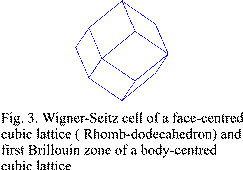
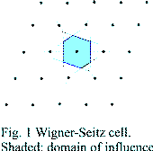
| 12:21, 25 January 2006 | Wigner3.gif (file) |  |
2 KB | 1 | |
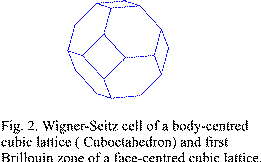
| 12:21, 25 January 2006 | Wigner2.gif (file) |  |
2 KB | 1 | |
| 12:21, 25 January 2006 | Wigner1.gif (file) |  |
1 KB | 1 | |
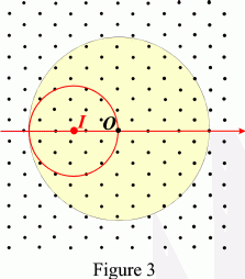
| 11:46, 25 January 2006 | Ewald-3.gif (file) |  |
7 KB | 1 | |
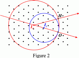
| 11:46, 25 January 2006 | Ewald-2.gif (file) |  |
5 KB | 1 | |
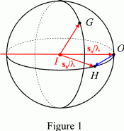
| 11:46, 25 January 2006 | Ewald-1.gif (file) |  |
4 KB | 1 |